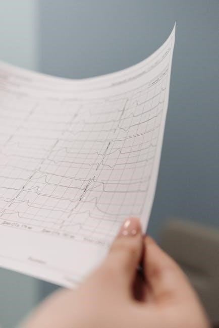
Rapid ECG interpretation is critical in emergency settings, enabling quick diagnosis and treatment․ It ensures accurate identification of life-threatening conditions like myocardial infarction and arrhythmias, improving patient outcomes․
Importance of Rapid ECG Interpretation in Emergency Settings
Importance of Rapid ECG Interpretation in Emergency Settings
In emergency situations, rapid ECG interpretation is vital for timely diagnosis and treatment․ It enables healthcare providers to quickly identify life-threatening conditions such as myocardial infarction, arrhythmias, and cardiac ischemia․ Accurate and swift interpretation ensures appropriate interventions, improving patient outcomes and reducing mortality․ ECGs provide critical insights into heart function, allowing clinicians to detect subtle abnormalities that may indicate serious underlying issues․ Delayed interpretation can lead to prolonged suffering or complications, while rapid analysis facilitates immediate decision-making․ This skill is particularly crucial in high-stakes environments like emergency rooms, where every second counts․ By mastering rapid ECG interpretation, healthcare professionals can deliver more efficient and effective care, ultimately saving lives․
Benefits of Using a Systematic Approach for ECG Reading
Using a systematic approach for ECG reading enhances accuracy and consistency, reducing errors and improving diagnostic confidence․ By following a structured method—such as assessing rate, rhythm, axis, and waveforms—clinicians can ensure no critical details are overlooked․ This approach minimizes missed diagnoses and allows for quick identification of abnormalities, such as arrhythmias or ischemia․ It also helps in organizing findings logically, making it easier to communicate with colleagues․ A systematic method is especially valuable in high-pressure environments, where time is limited․ Over time, this approach becomes second nature, speeding up interpretation while maintaining precision․ It also serves as a strong foundation for mastering advanced techniques and staying updated with continuous learning in ECG interpretation․

Basic Components of an ECG
An ECG is composed of distinct waveforms: P-wave (atrial depolarization), QRS complex (ventricular depolarization), T-wave (ventricular repolarization), and occasionally a U-wave, reflecting cardiac electrical activity in sequence․
Patient Details and Situation Assessment
Patient Details and Situation Assessment
Assessing patient details and the clinical situation is the first step in rapid ECG interpretation․ This includes the patient’s age, gender, medical history, and current symptoms․ Knowing the context helps identify patterns that may indicate underlying conditions․ For example, chest pain in a middle-aged patient with a history of hypertension could suggest ischemia․ The ECG should be interpreted alongside vital signs and physical examination findings to guide immediate management․ Accurate patient details ensure that abnormalities are not overlooked, enabling timely diagnosis and treatment․ This systematic approach enhances the reliability of ECG interpretation, especially in emergency settings where seconds count․
Measuring the Heart Rate on an ECG
Measuring the Heart Rate on an ECG
Measuring the heart rate on an ECG is a fundamental step in rapid interpretation․ For regular rhythms, count the number of complexes in a 6-second strip and multiply by 10 to estimate beats per minute (bpm)․ Alternatively, use the formula 300 divided by the number of complexes within a 5-second strip․ For irregular rhythms, measure the average R-R interval and calculate 60 divided by the interval in seconds․ Accurate heart rate assessment is crucial for identifying bradycardia (<60 bpm) or tachycardia (>100 bpm), which are key to diagnosing conditions like atrial fibrillation or sinus tachycardia․ This step ensures proper triage and treatment in emergency settings, where precise measurements are critical for patient care․

Assessing the Rhythm on an ECG
Assessing the Rhythm on an ECG
Assessing the rhythm on an ECG involves evaluating the pattern of heartbeats to determine if they are regular or irregular, which is crucial for identifying conditions like atrial fibrillation․ Identify P waves and their relationship with QRS complexes; consistent P waves preceding each QRS suggest a sinus rhythm, while their absence may indicate ventricular arrhythmias․ Measure the PR interval to detect abnormalities such as pre-excitation or heart block․ Examine QRS complex width to differentiate between supraventricular and ventricular arrhythmias․ Assess the rate to identify tachycardia or bradycardia, aiding in diagnosing conditions like SVT or sinus tachycardia․ Note ectopic beats, which can indicate PVCs or APCs․ Evaluate rhythm stability and patterns suggesting AV blocks or junctional rhythms․ Quick and accurate rhythm assessment is vital for timely clinical decisions and patient outcomes in emergency settings․

Understanding ECG Waveforms
ECG waveforms include P, Q, R, S, T, and U waves, each representing specific cardiac electrical activities․ The P wave signifies atrial depolarization, while the QRS complex marks ventricular depolarization, the ST segment reflects ventricular repolarization, and the T wave indicates ventricular recovery․ U waves are less common but may appear post-T wave, often linked to ventricular repolarization․ Accurately interpreting these waveforms is essential for diagnosing arrhythmias, ischemia, or other cardiac conditions, enabling timely clinical interventions․
P-Wave and PR Interval Interpretation
The P-wave represents atrial depolarization, with normal duration of 80-100ms and amplitude ≤2․5mm․ A absent or inverted P-wave may indicate atrial fibrillation or other arrhythmias․ The PR interval measures the time from P-wave onset to QRS complex, reflecting AV nodal conduction․ Normal PR ranges from 120-200ms; prolongation suggests first-degree AV block, while shortening may indicate pre-excitation syndromes like Wolff-Parkinson-White․ Accurate interpretation of these components is crucial for identifying conduction abnormalities and atrial pathology․ Changes in P-wave morphology or PR interval duration can signal conditions like atrial enlargement or accessory pathways, emphasizing the importance of systematic analysis in rapid ECG interpretation to guide timely clinical decisions․
Q-Wave and QRS Complex Analysis
The QRS complex is the most prominent part of the ECG, representing ventricular depolarization․ A normal QRS duration is ≤120ms, with the Q-wave being the first downward deflection․ Q-waves are abnormal if they are >40ms in duration or ≥25% of the QRS amplitude․ Pathological Q-waves suggest myocardial infarction or scarring․ The R-wave represents ventricular muscle depolarization, while the S-wave is the downward deflection following the R-wave․ Analyzing the QRS complex helps identify conditions like bundle branch blocks or ventricular hypertrophy․ Q-wave abnormalities are critical in diagnosing ischemic events, making their accurate interpretation essential for rapid ECG analysis and timely clinical intervention․
ST Segment and T-Wave Interpretation
The ST segment and T-wave are critical components of the ECG, reflecting ventricular repolarization․ The ST segment is the flat line following the QRS complex, while the T-wave represents ventricular muscle repolarization․ Normally, the ST segment is isoelectric or slightly elevated, and the T-wave is upright in most leads․ ST segment elevation or depression indicates myocardial ischemia or infarction, while T-wave inversion suggests ischemia, ventricular hypertrophy, or other pathologies․ Accurate interpretation of these waveforms is vital for diagnosing conditions like acute coronary syndromes or electrolyte imbalances․ Proper analysis requires comparing these segments across multiple leads to identify patterns and localize abnormalities, ensuring timely and appropriate clinical interventions․

Advanced ECG Interpretation Techniques
Advanced techniques involve calculating heart rate in irregular rhythms, assessing the axis, and identifying U waves․ These skills enhance diagnostic accuracy and clinical decision-making in complex cases․
Calculating Heart Rate in Irregular Rhythms
Calculating Heart Rate in Irregular Rhythms

In irregular rhythms, heart rate calculation requires alternative methods․ One approach is to count the number of R-R intervals over a 6-second strip and extrapolate to beats per minute․ For example, if 8 R waves occur in 6 seconds, the rate is approximately 80 beats per minute․ Another method involves measuring the average interval over 10 seconds to improve accuracy․ Irregular rhythms, such as atrial fibrillation, often require this technique․ Accurate calculation is crucial for diagnosing tachycardia or bradycardia․ Small errors in measurement can lead to significant misinterpretations․ Using these systematic methods ensures precise heart rate determination, even in complex arrhythmias․
Assessing the Axis on an ECG
Assessing the Axis on an ECG
Assessing the axis on an ECG involves determining the direction of the heart’s electrical activity․ The normal axis ranges from 0 to 90 degrees․ To determine the axis, examine the QRS complexes in leads I, II, and III․ Measure their amplitudes to identify the overall direction of electrical activity․ If the QRS in lead I is positive and in lead III is negative, it may indicate a specific axis deviation․ Left axis deviation (below 0 or above 180 degrees) is often linked to conditions like left ventricular hypertrophy, while right axis deviation (90 to 180 degrees) may indicate right ventricular hypertrophy․ Using systematic methods and reference guides can help accurately determine the axis, crucial for diagnosing heart conditions․ Practicing with real ECG examples enhances understanding and interpretation skills․
Identifying U Waves and Their Clinical Significance
Identifying U Waves and Their Clinical Significance
U waves are small, rounded deflections that appear after the T wave on an ECG, typically in the same direction as the T wave․ They are best observed in leads V2-V4 and are often associated with hypokalemia or hypomagnesemia․ U waves may also indicate myocardial ischemia or drug effects, such as with digoxin or quinidine․ To identify U waves, measure their amplitude and ensure they are distinct from the T wave․ Their clinical significance lies in their association with electrolyte imbalances and cardiac conditions like heart failure or arrhythmias․ Accurate identification and interpretation of U waves are crucial for diagnosing underlying pathologies, emphasizing the importance of context and systematic ECG analysis․

Common ECG Patterns and Their Meanings
Common ECG patterns include normal sinus rhythm, ST elevation, Q-waves, bundle branch blocks, and arrhythmias like atrial fibrillation․ Recognizing these patterns helps diagnose conditions quickly and accurately․
Recognizing Normal and Abnormal ECG Patterns
Recognizing Normal and Abnormal ECG Patterns
A normal ECG displays a consistent P-wave, QRS complex, and T-wave, with a regular rhythm and normal intervals․ Abnormal patterns include ST-segment elevation, Q-waves, or irregular rhythms, indicating conditions like myocardial infarction or arrhythmias․ Systematic analysis helps differentiate between benign variations and pathological changes․ For example, ST depression may suggest ischemia, while wide QRS complexes could indicate bundle branch blocks․ Recognizing these patterns quickly is essential for timely interventions․ Resources like ECG cheat sheets and step-by-step guides provide clear criteria for distinguishing normal from abnormal findings, ensuring accurate and rapid interpretation in clinical settings․ Regular practice with real-world examples enhances proficiency in identifying these critical patterns․
Identifying Life-Threatening Arrhythmias
Identifying Life-Threatening Arrhythmias
Identifying life-threatening arrhythmias on an ECG is crucial for immediate intervention․ Ventricular fibrillation (VF) and ventricular tachycardia (VT) are chaotic rhythms with no discernible P-waves and wide QRS complexes․ Atrial fibrillation (AF) shows an irregular, quivering baseline with no P-waves․ Supraventricular tachycardia (SVT) presents as a rapid, regular rhythm with a narrow QRS․ Torsades de Pointes is characterized by a twisting QRS axis and prolonged QT interval․ These arrhythmias require urgent treatment, such as defibrillation or medication․ Systematic analysis using ECG cheat sheets and real-world examples helps in rapid recognition․ Early identification ensures timely interventions, improving patient outcomes in emergencies․ Regular practice with clinical cases enhances the ability to quickly diagnose these critical conditions․

Practical Application of ECG Interpretation
Practical application involves using clinical cases, real-world examples, and self-study materials to enhance diagnostic skills․ Quick reference guides and step-by-step approaches improve accuracy and patient care․
Clinical Cases and Real-World Examples
Clinical Cases and Real-World Examples
Clinical cases and real-world examples are essential for mastering rapid ECG interpretation․ They provide practical insights into recognizing patterns and applying systematic approaches․ Real patient scenarios, such as STEMIs, arrhythmias, and conduction disorders, help learners identify abnormalities and correlate findings with symptoms․ ECG libraries, like LITFL ECG Library, offer high-quality images and detailed interpretations, enabling users to practice and refine their skills․ Step-by-step guides and clinical blogs further enhance understanding by breaking down complex cases․ These resources emphasize contextual learning, ensuring that interpretations are accurate and clinically relevant․ By studying diverse examples, learners can improve their ability to diagnose and manage cardiac conditions effectively, ultimately improving patient outcomes․
Using ECG Examples for Self-Study and Practice
Using ECG Examples for Self-Study and Practice
Using ECG examples for self-study and practice is a highly effective way to master rapid interpretation skills․ ECG libraries, such as those from LITFL, provide high-quality ECG images with detailed explanations, allowing learners to practice interpreting various patterns․ Quizzes and interactive tools further enhance learning by testing knowledge and reinforcing key concepts․ ECG examples cover a wide range of conditions, from normal patterns to life-threatening arrhythmias, ensuring comprehensive understanding․ Systematic approaches, as outlined in many guides, help users methodically analyze each case, improving accuracy and speed․ By regularly reviewing and practicing with real-world ECGs, individuals can refine their skills and build confidence in their ability to interpret ECGs effectively․

Resources for Rapid ECG Learning
ECG libraries, cheat sheets, and online tools provide essential resources for rapid learning․ Step-by-step guides and real-world examples enhance understanding and improve interpretation skills effectively․
ECG Cheat Sheets and Quick Reference Guides

ECG Cheat Sheets and Quick Reference Guides
ECG cheat sheets and quick reference guides are invaluable tools for rapid interpretation․ They provide concise, step-by-step algorithms for identifying key waveforms, intervals, and patterns․ These resources often include visual aids, such as diagrams of P-waves, QRS complexes, and ST segments, to help learners quickly recognize abnormalities․ Many guides also highlight critical conditions like myocardial infarction or life-threatening arrhythmias․ Online platforms offer downloadable PDFs, such as Dr․ Martin Milner’s one-page ECG interpretation guide, which simplifies complex concepts․ These tools are particularly useful for students and professionals in emergency settings, enabling them to make accurate and timely decisions․ By focusing on essential elements, cheat sheets streamline the learning process and enhance proficiency in rapid ECG interpretation․
Recommended Online Tools for ECG Interpretation
Recommended Online Tools for ECG Interpretation
Several online tools and resources are available to enhance rapid ECG interpretation skills․ Websites like ECGwaves․com and Litfl․com offer comprehensive libraries of ECG examples, clinical cases, and step-by-step guides․ These platforms provide interactive tools for practicing interpretation and include high-quality images for download․ Additionally, resources like ECG Guide & Quiz and Dr․ Martin Milner’s ECG interpretation guide are widely recommended․ These tools often include algorithms, diagrams, and real-world examples to simplify learning․ Many platforms also offer downloadable PDF guides and mobile-friendly interfaces for quick reference․ These resources are particularly beneficial for students and professionals in emergency settings, enabling them to refine their skills and improve diagnostic accuracy․ By leveraging these tools, learners can master rapid ECG interpretation efficiently․
Step-by-Step Guides for Beginners
Step-by-Step Guides for Beginners
Step-by-step guides are essential for mastering rapid ECG interpretation, especially for beginners․ Resources like Dr․ Martin Milner’s 1-page ECG interpretation guide provide a concise approach to learning․ These guides break down the process into manageable steps, such as assessing patient details, measuring heart rate, and analyzing waveforms․ Many online platforms, including ECGwaves․com and Litfl․com, offer structured learning pathways with real-world examples and interactive exercises․ These guides often include algorithms, diagrams, and clinical cases to simplify complex concepts․ Additionally, downloadable PDF guides and mobile-friendly tools allow learners to practice anytime, anywhere․ By following these step-by-step resources, beginners can build a strong foundation in ECG interpretation and improve their diagnostic skills efficiently․
Mastery of rapid ECG interpretation requires dedication and practice․ Systematic approaches and continuous learning ensure accurate diagnoses, improving clinical decision-making and patient outcomes in emergency settings․
Final Tips for Mastering Rapid ECG Interpretation
Final Tips for Mastering Rapid ECG Interpretation
To excel in rapid ECG interpretation, start by mastering the basics, such as recognizing normal and abnormal waveforms․ Always use a systematic approach, beginning with patient details and moving through rate, rhythm, axis, and waveform analysis․ Regular practice with real-world examples and self-study tools sharpens skills․ Utilize cheat sheets and quick reference guides to reinforce key concepts․ Focus on identifying life-threatening patterns, such as ST-segment elevations or wide QRS complexes, which demand urgent action․ Continuous learning through clinical cases and updated resources ensures proficiency․ Finally, correlate ECG findings with clinical context to enhance diagnostic accuracy and patient care․
The Role of Continuous Learning in ECG Proficiency
Continuous learning is essential for mastering rapid ECG interpretation․ Regular engagement with clinical cases, updated guides, and online resources ensures proficiency․ Systematic approaches, like those in ECG interpretation guides, help refine skills․ Practicing with real-world examples and self-study tools enhances accuracy․ Staying informed about new patterns and advancements keeps interpretations current․ Engaging with communities and experts fosters a deeper understanding․ Learning from mistakes and feedback improves diagnostic precision․ Ultimately, continuous learning ensures clinicians remain adept at identifying critical conditions, providing timely care․
